

Dental caries, commonly known as tooth decay, is a prevalent oral health issue affecting people of all ages. To effectively diagnose and treat caries, dental professionals often rely on standardized classification systems. One such system is the G.V. Black Classification of caries, developed by Dr. Greene Vardiman Black, a prominent American dentist, in the late 19th century. In this blog post, we'll delve into the G.V. Black Classification system, its significance, and how it helps dentists in diagnosing and treating dental caries.
The Origins of G.V. Black Classification
The Origins of G.V. Black Classification
Dr. G.V. Black is often referred to as the "father of modern dentistry" for his pioneering work in the field. He recognized the need for a standardized system to categorize dental caries, as it would facilitate communication among dental professionals and improve the overall quality of dental care. Thus, he developed the G.V. Black Classification system, which is still widely used today.
The G.V. Black Classification System
The G.V. Black Classification System
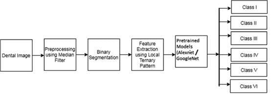
The G.V. Black Classification system categorizes dental caries based on their location, extent, and severity. It utilizes a simple alphanumeric system to describe each type of caries lesion, making it easy for dentists to communicate and record the condition of a patient's teeth.
The Importance of Precise Categorization
The Importance of Precise Categorization
The G.V. Black Classification system is more than just a tool for communication among dental professionals; it plays a pivotal role in the precise diagnosis and treatment of dental caries. When a dentist encounters a patient with carious lesions, they must accurately categorize them to provide the most appropriate care. Let's explore how this system is applied in practice.
Class I - Occlusal Caries
Class I - Occlusal Caries
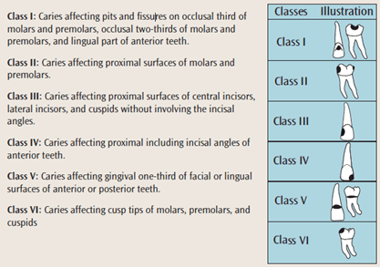
Class I caries are typically found on the chewing surfaces of molars and premolars, as well as in the occlusal pits of posterior teeth. These lesions often result from the accumulation of food particles and bacteria in the deep fissures and grooves of these teeth. When diagnosing Class I caries, dentists can use various diagnostic tools such as visual examination, probing, and dental X-rays. Once identified, the treatment for Class I caries usually involves dental fillings, sealants, or other restorative procedures, depending on the extent of the decay.
Class II - Proximal Caries
Class II - Proximal Caries
Class II caries primarily affect the proximal surfaces, which are the sides that touch adjacent teeth. These interproximal caries are often challenging to diagnose without the aid of dental X-rays. Early detection is crucial to prevent the progression of the caries, which could lead to more extensive and costly treatments like dental crowns or root canals.
Class III - Proximal Caries in Anterior Teeth
Class III - Proximal Caries in Anterior Teeth
Class III caries are similar to Class II but are specific to anterior teeth, including incisors and canines. Diagnosing these caries involves examining the proximal surfaces, and the treatment typically includes dental fillings or bonding to restore the damaged tooth structure.
Class IV - Proximal Caries in Anterior Teeth with Incisal Edge
Class IV - Proximal Caries in Anterior Teeth with Incisal Edge
Class IV caries encompass not only the proximal surfaces but also extend to include the incisal (biting) edge of anterior teeth. These are often the most noticeable caries lesions because they can significantly affect the tooth's appearance. Treating Class IV caries often involves more extensive restorations to ensure both the functional and aesthetic aspects of the tooth are restored.
Class V - Gingival Caries
Class V - Gingival Caries
Class V caries affect the gingival (gum) one-third of the facial or lingual surfaces of any tooth. These caries are often associated with gum recession, making them more susceptible to decay. Treatment involves dental fillings or other restorative procedures to protect the exposed root surface and prevent further decay.
Class VI - Incisal or Occlusal Caries
Class VI - Incisal or Occlusal Caries
Class VI caries occur on the incisal or occlusal edges of teeth. These caries often result from wear and tear over time. Treatment varies depending on the extent of the damage and may involve dental fillings, inlays, onlays, or crowns.
Contemporary Tools and Techniques
Contemporary Tools and Techniques
Digital radiography provides high-resolution images with lower radiation exposure, enhancing the detection of caries, even in their early stages. Intraoral cameras allow dentists to capture images of carious lesions, providing a visual record for both diagnosis and patient education.
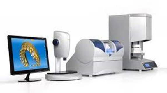
CAD/CAM technology has revolutionized the process of creating dental restorations. Dentists can design and fabricate crowns, inlays, onlays, and other restorations in a single appointment, offering patients more convenience and expedited treatment.
Conclusion
The G.V. Black Classification of Dental Caries remains the cornerstone of caries diagnosis and treatment, ensuring that dental professionals can accurately categorize and address each patient's unique needs. As the field of dentistry continues to advance, new technologies and techniques further enhance the precision and efficiency of caries diagnosis and treatment. This ongoing evolution, combined with the foundational principles of the G.V. Black Classification, continues to benefit patients by preserving their oral health and beautiful smiles.
